Research
We want to understand how a brain builds internal representations of the world and of the animal's own actions, and how it uses them to behave. We work mostly in the larval zebrafish, a small transparent vertebrate in which the activity of every neuron in the brain can be imaged while the animal behaves, and we combine whole-brain imaging with behavior, anatomy and theory. We have recently started to do experiments in mice as well, to ask which of the principles we find in the fish hold across vertebrates.
An internal compass
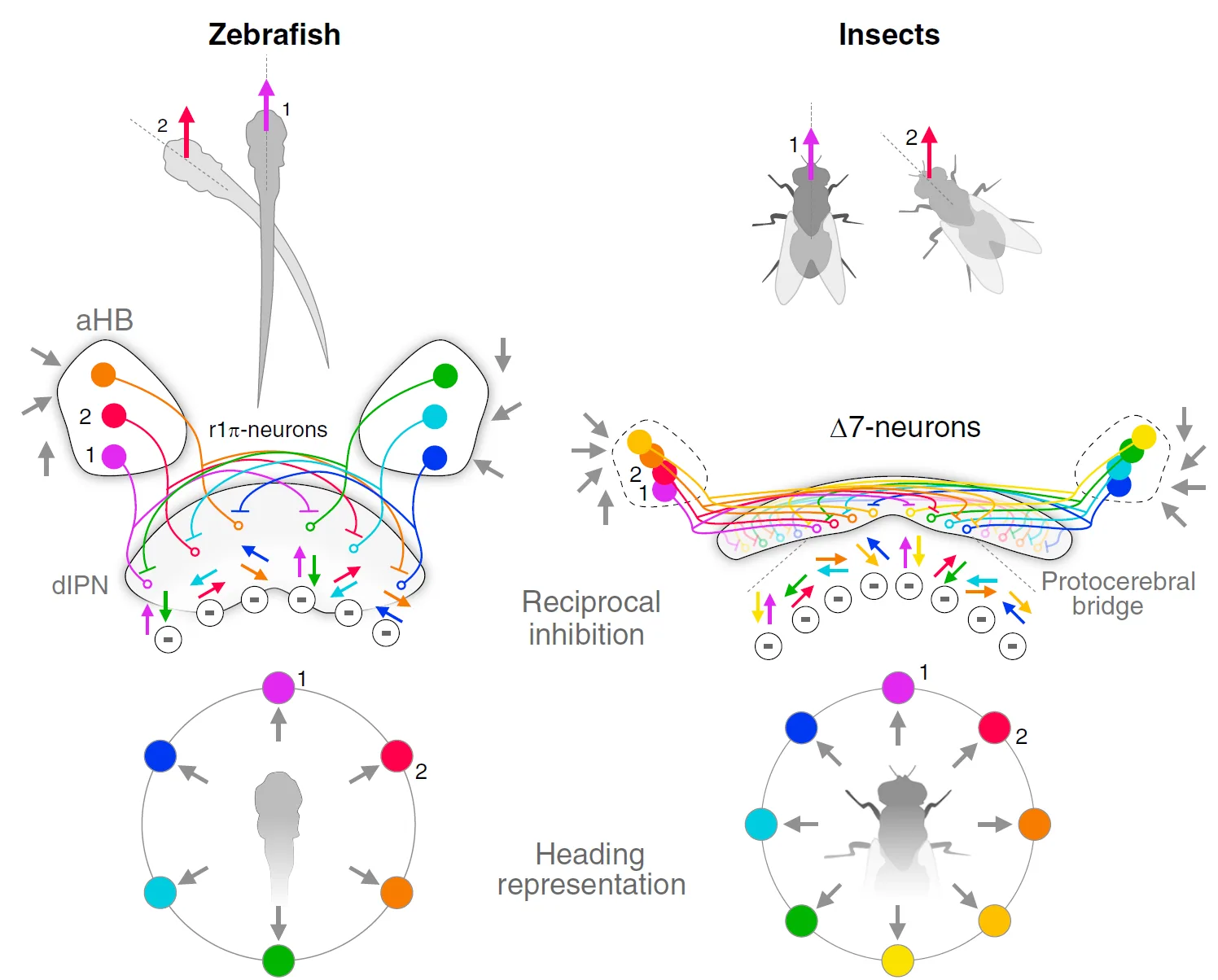
Larval zebrafish carry a compass in their brain. A population of neurons in the anterior hindbrain, projecting to the interpeduncular nucleus (IPN), holds a single bump of activity whose position tracks the direction the fish is facing. The bump persists in darkness, moves when the animal turns, and is stable when the animal is still: the hallmarks of a ring attractor network. In its simplest description, the network integrates the animal's angular velocity ω, so that the bump position θ(t) = θ0 + ∫ω dt keeps track of heading even without any sensory cue.
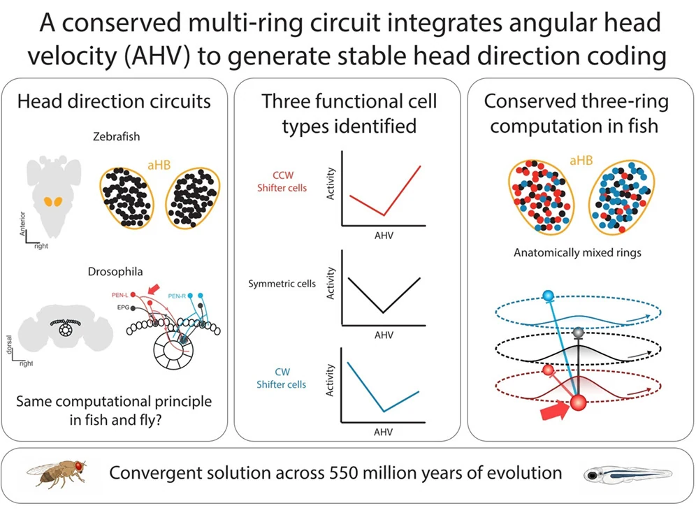
We are working out how this computation is implemented. By fitting network models to recordings, we found that the fish compass, like the one in the fly, is built from a main ring and two shifter rings that push the bump clockwise or counterclockwise, even though the three populations are anatomically intermingled. We are also studying how the compass is tied to the world: motion of the visual scene and the position of landmarks reach the IPN and align with heading, and the mapping between a landmark and a heading is plastic, so that the compass can be re-anchored when the world changes.
Key papers: Petrucco et al., Nature Neuroscience 2023 (heading direction neurons and ring attractor dynamics in the fish hindbrain) · Lavian et al., Nature Communications 2025 (visual motion and landmark inputs to the IPN) · Tanaka & Portugues, Nature 2026 (plastic landmark anchoring) · Mei et al., Current Biology 2026 (a multi-ring shifter network)
The compass circuit: anatomy, identity, and a second species
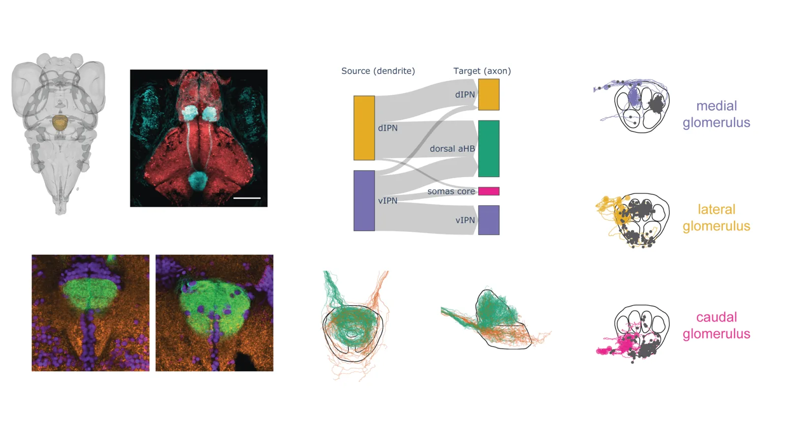
Knowing what a circuit computes is only half of the problem; we also want to know what it is made of. We are reconstructing the IPN and its inputs at synaptic resolution from electron microscopy, to test directly whether the connectivity contains the feedback loops with an angular offset that a shifter network requires. In parallel, we are characterising the molecular identity of the compass neurons, in order to generate genetic access to them and to ask whether they are homologous to the head direction cells of the mammalian brainstem.
The larval zebrafish gives us a compass we can see in its entirety. To ask which of its properties are general, we are starting to do experiments in mice, where the head direction system has been studied for decades but is far harder to observe as a whole. We bring the same questions to the mouse that we ask in the fish: what drives the bump, what anchors it to the world, and how the circuit that computes it is wired.
Learning which actions lead to which outcomes
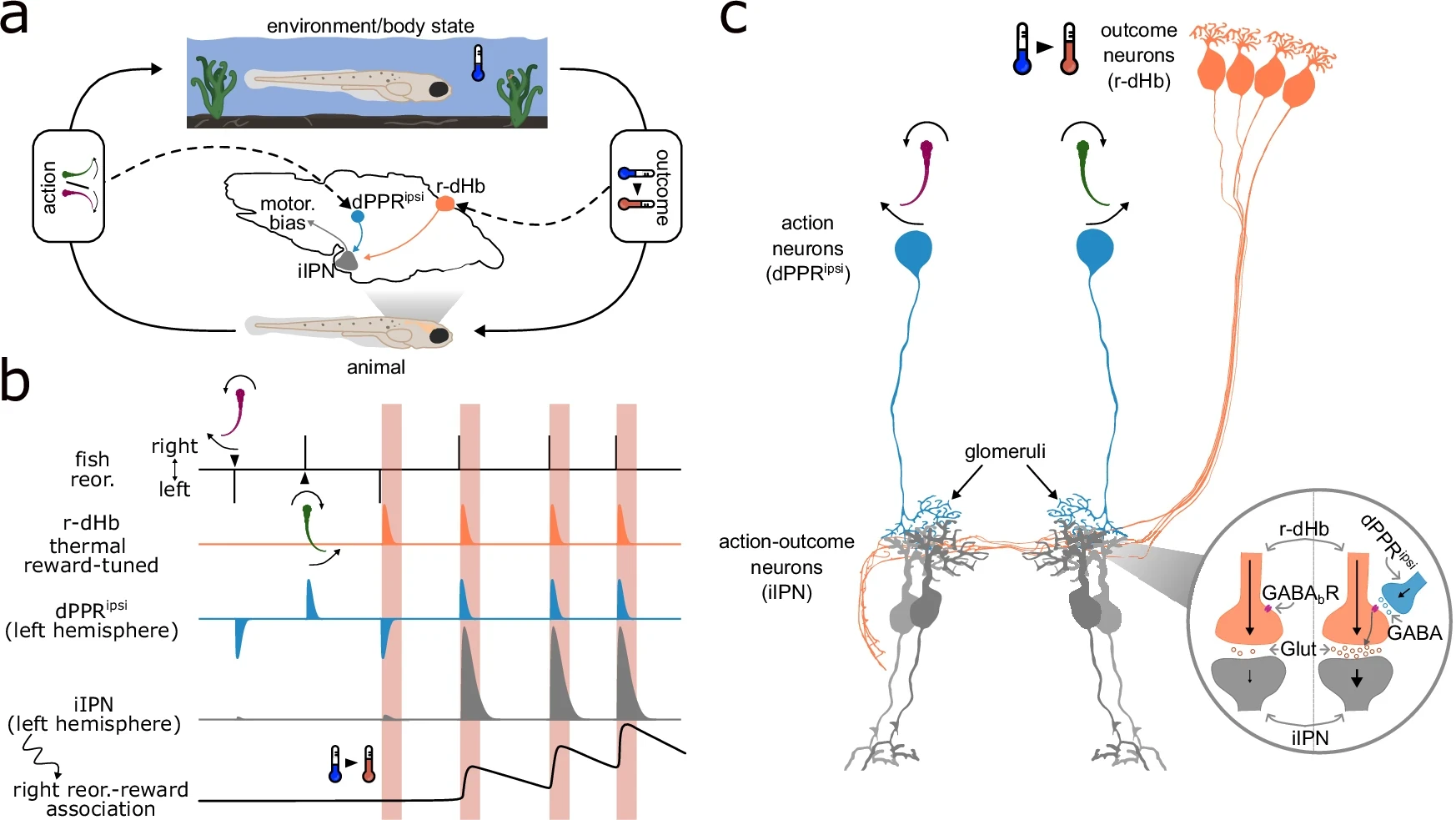
Improving with experience requires solving a deceptively simple problem: something good happens, and the animal has to work out which of the things it just did was responsible. A larval zebrafish placed in water that is too cold will learn, in a handful of trials, that turning in one particular direction warms it up, and it will do so only if the warmth follows the turn within a fraction of a second.
We found the circuit that solves this problem. The habenula carries the outcome ("I got warmer"), a small group of inhibitory hindbrain neurons carries the action ("I turned left"), and the two signals meet on the habenular axon terminals inside a single glomerulus of the IPN. There, presynaptic GABAB receptors do something unusual: instead of dampening transmitter release, they boost it, so that only an action followed by its outcome drives the postsynaptic neurons. Blocking these receptors abolishes both the neural response and the learning. The dorsal habenula, together with the preoptic area, also supports the slower homeostatic navigation that keeps the fish near the temperatures it prefers. We are now asking how these thermal, navigational and compass signals interact within the IPN.
Key papers: Palieri et al., Current Biology 2024 (preoptic area and habenula in homeostatic navigation) · Paoli et al., Nature Communications 2026 (action-outcome association at habenular terminals)
Internal models and the sensory consequences of movement
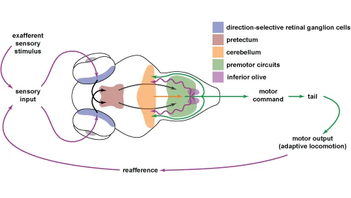
Every movement changes what an animal sees and feels. A swimming fish sees the world slide backwards and feels water flow along its body, and it has to distinguish these self-generated signals from the ones that carry news about the world. Much of our earlier work asked how the brain does this and how it adapts when the relationship between action and consequence changes.
Using whole-brain imaging during behavior, we described the distributed networks that turn visual motion into swimming, showed that the brain accumulates evidence about the direction of motion before committing to a turn, and found that the cerebellum maintains an internal model of the consequences of swimming: when visual feedback is persistently altered, this model is updated and recalibrates a fast feedback controller so that the fish swims appropriately for the new conditions. We have also asked how visual motion is remembered over seconds, and how the lateral line distinguishes self-generated flow from that of the environment.
Key papers: Ahrens et al., Nature 2012 (brain-wide dynamics during motor adaptation) · Portugues et al., Neuron 2014 (whole-brain maps of visuomotor behavior) · Dragomir et al., Nature Neuroscience 2020 (evidence accumulation) · Markov et al., Nature Communications 2021 (a cerebellar internal model) · Odstrcil et al., Current Biology 2022 (reafferent mechanosensation) · Tanaka & Portugues, Current Biology 2025 (optic flow memory)
Fish, flies and the theory of neural computation
Fish and flies last shared an ancestor around 550 million years ago, yet both carry a ring attractor compass, both anchor it to visual landmarks, and both use modulation of axon terminals to assign credit to actions. We find these parallels irresistible. Part of our work is explicitly comparative, drawing on the beautifully detailed picture of the insect brain to generate hypotheses for the vertebrate one, and vice versa.
Underlying all of this is a commitment to theory. We build network models of the circuits we record from, use them to ask which architectures can and cannot produce the dynamics we observe, and think about heading estimation as a problem of integration and inference under uncertainty. The aim is to arrive at descriptions of neural computation that are precise enough to be wrong.
Key papers: Tanaka & Portugues, Nature Reviews Neuroscience 2025 (analogies between vertebrate and insect visual systems) · Mei et al., Current Biology 2026 (the same ring attractor architecture in fish and fly)
How we work
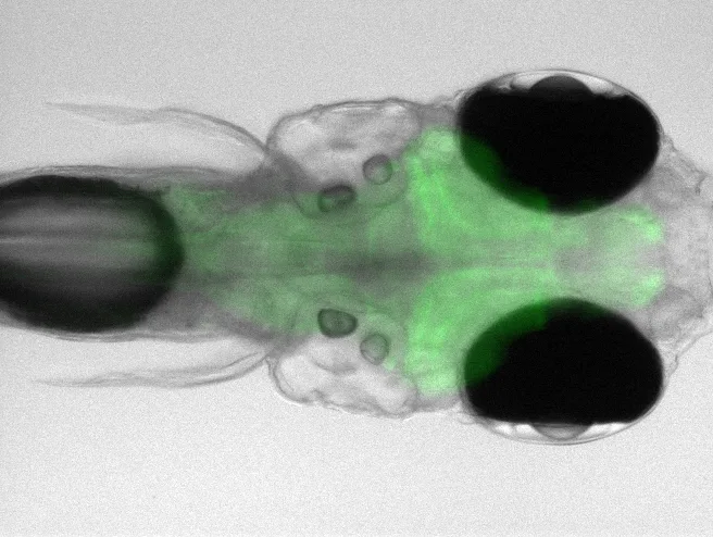
Our experiments rest on imaging the whole brain of a behaving larval zebrafish at cellular resolution, with lightsheet and two-photon microscopy, while the animal navigates virtual environments that we control in closed loop. We build our own rigs and write our own software, and we release it: Stytra, our system for stimulation, tracking and closed-loop experiments, and our contributions to BrainGlobe and brainrender for anatomical registration and visualisation, are used by many labs. Alongside imaging we use electron microscopy, transgenics and spatial transcriptomics to describe the circuits anatomically and molecularly, and network modelling to make sense of what we record.
Tools: Štih et al., PLOS Computational Biology 2019 (Stytra) · Claudi et al., JOSS 2020 (BrainGlobe Atlas API) · Claudi et al., eLife 2021 (brainrender)
We are always looking for curious people who want to work at the interface of experiment and theory, whatever their background. If any of this sounds like the kind of problem you would like to spend a few years on, get in touch.